Doctors Are Able to Make Sonograms Through the Use of
You may also hear the word sonography used in reference to the ultrasound exam. The speed of sound depends on.

Pin On Baby Toddler
Ultrasound probes called transducers produce sound waves that have frequencies above the threshold of human hearing above 20KHz but most transducers in current use operate at much higher frequencies in the megahertz MHz range.
. The technique relies on high-frequency sounds to create the picture. Many people associate this procedure with the abdominal ultrasounds performed during pregnancy. However recent advancements in technology have created ultrasound imaging that is three-dimensional 3-D.
Your doctor uses both to complete the procedure. Doctors can use ultrasound results to diagnose a wide range of conditions. The images produced from the sound waves can help experts get a clear visual of what is happening inside the body.
Sonogram is used for various purposes and is also helpful in checking the heart rate and pregnancy status of woman. A sonogram may be used to identify the gender of an unborn baby. All this information allows the physician to assess and treat.
But gradually with developing expertise they could discern fine structures in. 13 Kidney Function. This technology which is used in different types of sonograms is extremely helpful in judging the.
The sonogram is the image. Ultrasound machines in a doctors office that treat vascular issues allows for a close and quick view of blood as it flows through the body detect blood clots locate blockages and impediments determine if arteries are enlarged as well as determine the source of varicose veins. Conventional ultrasounds can display their images as thin flat sections of the body.
The pitch of a. A form of ultrasound known as Doppler imaging is used for creating the image which is able to make various calculations pertaining to the speed direction and volume of blood that flows into and out of the heart. Ultrasound is the technology used to create the image.
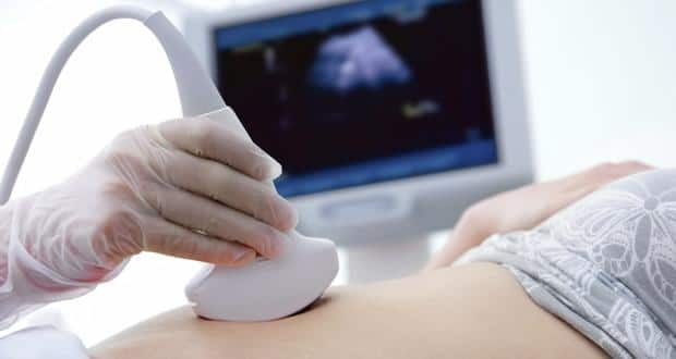
Thus during pregnancy doctors use ultrasound tests to examine the fetus inside a woman and are able to view the structure and any movement of the fetus. An ultrasound technician called a sonographer will apply a special lubricating jelly to your skin. Ultrasound imaging also known as sonography uses high-frequency sound waves to create images of the inside of the body.
This prevents friction so they can rub the ultrasound transducer on your skin. Over the next decade other doctors refined the procedure to detect breast tumors and to perform echocardiograms. Sometimes however doctors need images of internal structures of the body to make a diagnosis and come up with a treatment recommendation.
You have a baby to make. Karl Dussik of Austria is credited with being the first person to use ultrasound to try to detect tumors Dr. Discover the capabilities of ultrasound imaging and its safety.
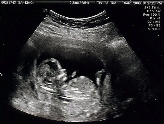
A sonogram is a picture of the internal organs of the body taken using the high precision machines which use high frequency or ultra sound waves. Doctors frequently are able to diagnose a medical issue by analyzing a list of symptoms and looking at a patient with only minimal equipment. During an ultrasound doctors will look at several of the major developing organs to ensure that they are growing and working well.
It allows your doctor to monitor your babys growth detect abnormalities track milestones home in on your due date. It is through the bouncing of these sound waves from the organs inside the body that a picture of the same is created. For most pregnant women sonograms are a regular part of prenatal medical care which provides future parents with the first glimpses of their baby.
Voluson E10 ultrasound machine features 3D-printing technology for the first time in the OBGYN sectorWith the help of it parents can determine which congenital defects their children need to deal with like cleft lips and extremities anomalies as well as abdominal wall defects. Healthcare practitioners commonly use ultrasound technology to investigate unknown causes of internal pain as well as to diagnose and monitor unusual growths such as tumors. Can Doctors Make 3D Print Of Ultrasound For Blind Mothers.
Ask all the questions you need to and do what you need to do to get as zen as you can. Enjoy this time as much as you can. Sonogram is painless and safe and hence used on a large scale by doctors.
Elasticity density and temperature. An ultrasound is a noninvasive procedure that doctors use to diagnose patients. In 30 years you want to be able to look back on this time fondly.
- If the doctor had huge concerns he would have done the ultrasound right away. Ultrasound is also known as sonography which is where the confusion lies. The amount of energy a sound wave carries per second through a unit area is its.
You need separate certificates in order to conduct Doppler imaging on adults children and fetuses. Doctors are able to make sonograms through the use of. When practitioners are looking at the fetuss kidneys and bladder they will be able to note if a babys kidneys look enlarged.
Doctors are able to make sonograms through the use of ULTRASOUNDUltrasound. Dont stress to much. Diagnostic ultrasound is a non-invasive diagnostic technique used to image inside the body.
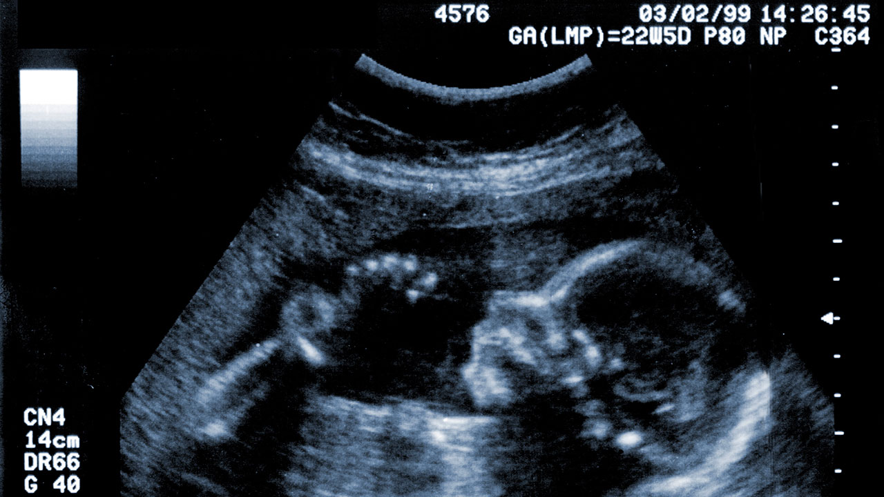
Sonography refers to the practice of using an ultrasound machine to create a sonogram and the sonographer is the skilled technologist who operates the equipment. The image or sonogram is generated through the process called ultrasonography. Early in the use of fetal ultrasound clinicians could only detect the babys head Nicolson said.
Ultrasound machines like the Philips IE33 allow technicians and doctors to see the inner structures and organs in a patient without having to make an incision. Thus theres very little difference between a sonogram vs. George Ludwig in the United States was the first to make a practical use of ultrasound to detect gallstones in 1948.
It is important to understand about the types of sonogram and its uses to make an informed decision.

Quick Facts Ultrasonography Msd Manual Consumer Version
Sonography The What How And When Of The Procedure Thehealthsite Com

Pin On Prion Diseases Spinal Muscular Atrophy Gene Therapy

Her Bun My Oven Baby Ultrasound Pictures Baby Ultrasound Ulzzang Kids

Pin On Momsense

3d Ultrasound Reveals Baby In Color Ultrasound Reveal 3d Ultrasound Ultrasound

Pin On Twin Sibling Connection
For Blind Moms 3 D Prints Of Fetuses Stand In For Sonogram Images Smart News Smithsonian Magazine

Pin On Theme 3 Societies And Government

Pin On U T

Pin On Theme 3 Societies And Government
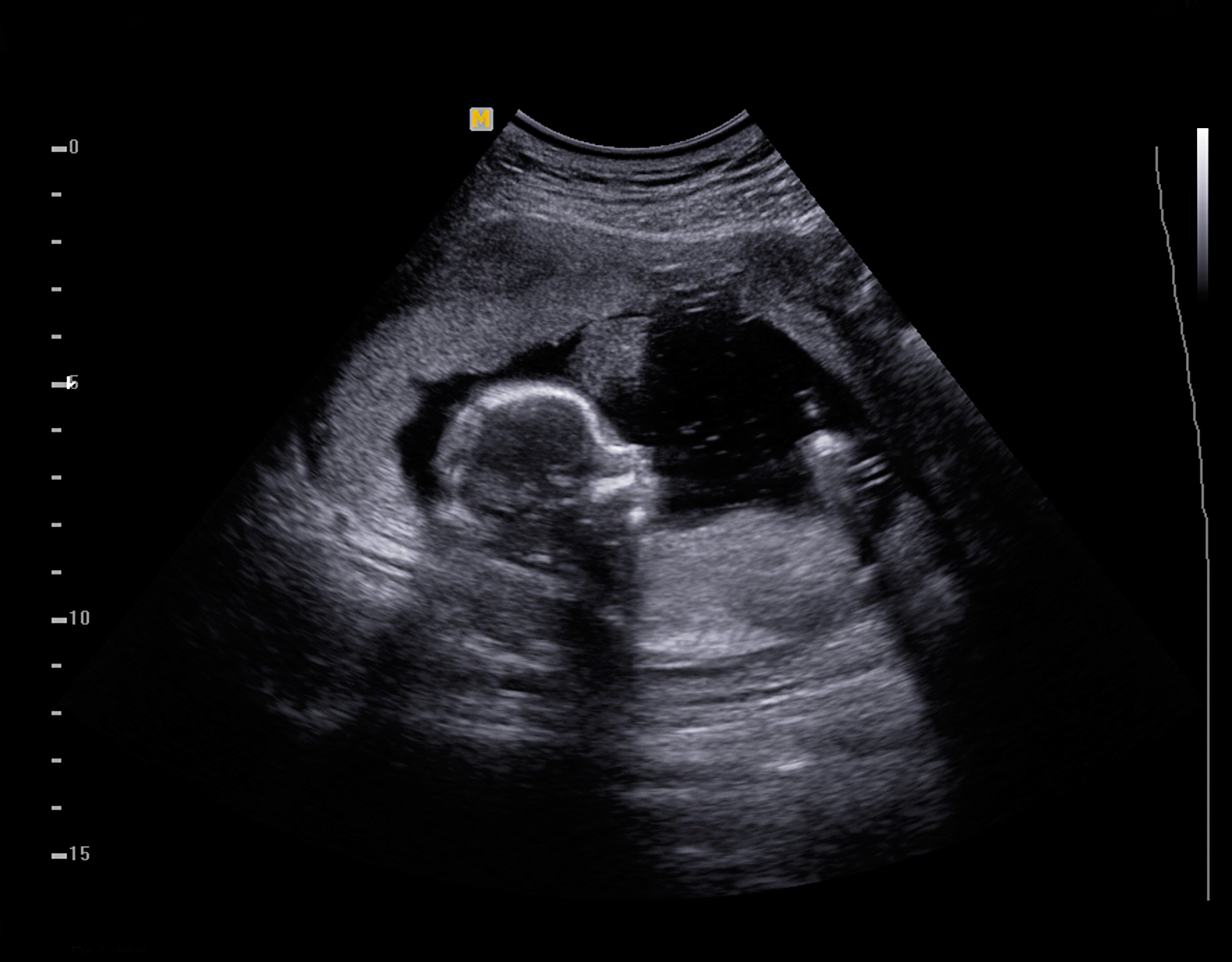
Pregnancy Dads The 20 Week Scan Raising Children Network

Pin On Projects To Try

Pin On Fake A Baby Pregnancy Gag Items
First Look At Your Baby The Fascinating History Of The Sonogram

Pin On Baby Stuff

Blue Damask Pattern Sonogram Baby Shower Invitation Zazzle Com Baby Shower Invitations Pink Baby Shower Invitations Animal Baby Shower Invitations

What Is Sonography And How Does It Work When It Comes To Sonography And Ultrasound What Is The Difference Focus Kimia
:focal(512x385:513x386)/https://tf-cmsv2-smithsonianmag-media.s3.amazonaws.com/filer_public/90/0b/900b6422-dbec-4369-a3e1-c21ca7bc8d5f/gettyimages-1219040940.jpg)
A Brief History Of The Sonogram Innovation Smithsonian Magazine


Comments
Post a Comment